Magnetic particle imaging, in short MPI, is an imaging technique that has been invented in 2005 by B. Gleich and J. Weizenecker.
The main area of application is medical imaging and related applications.
MPI is a tracer-based technique which is currently in the preclinical phase exploring potential clinical applications:
Magnetic nano-particles, each consisting of a magnetic iron-oxide core and a non-magnetic coating, could potentially be injected into the blood stream of a patient.
The particles are distributed inside the cardiovascular system.
By determining the resulting concentration of particles in the body, it is possible to draw conclusions on, e.g., cloggings in blood vessels, or to locate tumors.

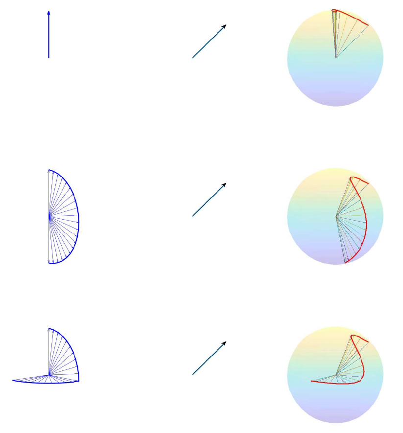
The basic idea is to exploit the response of the magnetic nano-particles to an external applied and highly dynamic magnetic field.
More specifically, a particle abruptly changes its magnetization if the external field changes rapidly in the vicinity of the position of this particle.
In order to encode information on the particles’ positions, the external magnetic field is designed in such a way that these rapid changes are limited to a small area, which is then driven through the field of view to scan the entire region of interest.
A common realization of this is a magnetic field with a field-free point (FFP).
If the FFP is moved through a position with a non-zero concentration of magnetic particles, it causes the magnetization of the particles to flip.
This abrupt change in the particle magnetization induces voltages in the receive coils of the scanner.
The goal in MPI is thus to reconstruct the concentration of the magnetic nano-particles inside a body from measurements of the voltages that are caused by temporal changes in the particle magnetization in response to the external magnetic field.
From an inverse problems point of view the imaging problem can be formulated by a Fredholm integral equation of the first kind for a given space-time-dependent integral kernel.
